Surgery Of The Future: Surgeons Using Printers To Make 3D Patient Models
Two surgeons who specialize in minimally invasive operations at Beth Israel Deaconess Medical Center have won a grant to use 3D printers to build models of patient body parts containing particular pathologies, such as tumors or aneurysms, that one day may be used to better educate patients about their illnesses and what will happen during their surgeries.
The 3D models will also be able to be used in the training of surgeons and may eventually serve to help surgeons better prepare for tricky operations by practicing first on the models.

"We are laying the groundwork and testing the waters," said Dr. Robert Andrews, who, along with Dr. Daniel Jones, received the $40,000 grant from the William F. Milton Fund of Harvard Medical School. The pair is working with Dr. Justin Kung, a radiologist, and Theodore Korelitz, a volunteer with an engineering background, to see just where the 3D models might take surgery in the future.
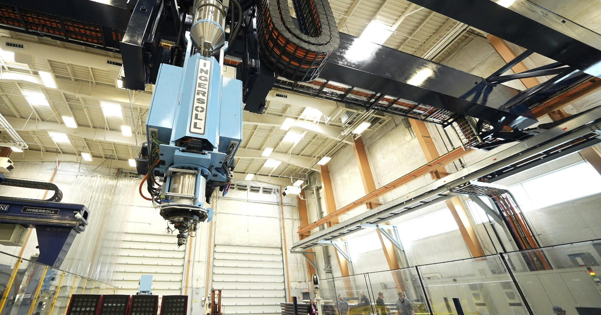
The models are created out of polymers using 3D printers made by Objet Geometries Ltd. The printers work by taking a 3D computer file, in these cases usually from a CT scan, and constructing from it a series of cross-sectional slices. Each slice is then printed, one on top of the other, to create the 3D model.
The models, for example, could be of a breast containing a tumor, or a liver with several tumors. Different polymers are used for different types of body tissues. The models are designed not unlike those created by architects designing buildings, for example.
"We're very lucky to have this grant," Dr. Andrews said. "Otherwise, we would not be making these models. They are very expensive."
Each model takes about 15 man hours of work to create.
Right now, the team is creating models from patient CT scans that are not identified by name since they are only being used for learning and testing the process, not in patient care.
Eventually, Dr. Andrews said, the models – which are uniquely created to be patient specific – would be able to be shown to the patients in question to help explain to them what is going on inside their bodies and what will happen during their surgeries.
"They can be used for better doctor-patient communication and in the informed consent process," he said.
Down the line, the models would likely be used in the training of doctors and one day by surgeons in the preparation of the operations to be performed on the patients who have been modeled. For example, if a surgeon needs to remove a tumor from a patient but there is a significant chance that he or she might hit a nerve or artery in doing so, the surgeon could create a model of the tumor from the patient's CT scans using the 3D printer. The surgeon could then practice on the model before working on the patient.
"When we go in there, we often find surprises," said Dr. Andrews. "Arteries and bile ducts, for example, in an operation as common as a gallbladder removal, can be highly variable from patient to patient. If we could model them in advance, it might very well help in knowing what steps or precautions to take in advance to avoid problems."
Above content provided by Beth Israel Deaconess Medical Center. For advice about your medical care, consult your doctor.
Posted: March 2013